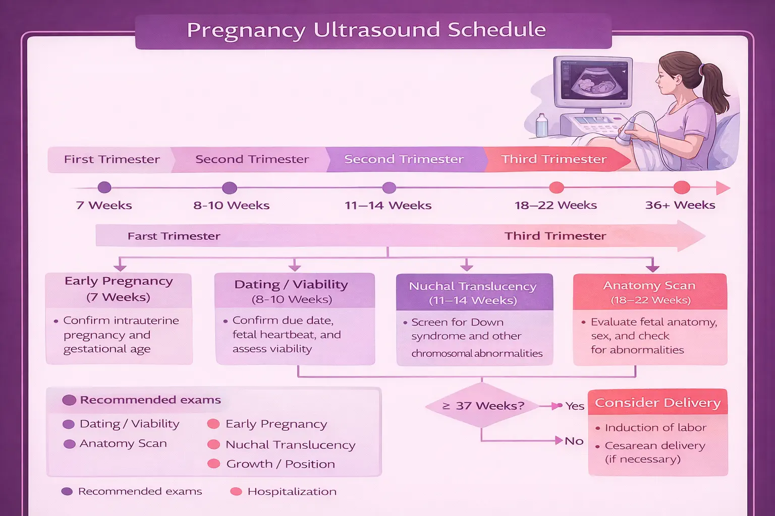
See your baby clearly. Diagnose with confidence.
Ultrasound is the primary imaging tool in obstetrics and gynecology — safe, radiation-free, and available in real time. At Broad Medical Group, Dr. Broad performs diagnostic and elective ultrasound in the office, reviewing results with you during your visit. This guide explains what each type of ultrasound shows, when scans are performed during pregnancy, and what to expect at your appointment.
Obstetric and gynecologic ultrasound uses sound waves to create real-time images of internal structures — no radiation is involved, and it is considered safe throughout pregnancy ACOG PB #175. 2D ultrasound is the standard diagnostic modality for pregnancy dating, anatomy surveys, growth monitoring, and gynecologic evaluation. 3D ultrasound adds surface rendering that is particularly useful for evaluating facial and skeletal abnormalities ISUOG 2020. 4D ultrasound provides real-time 3D video of fetal movement. Most low-risk pregnancies include at least a dating scan (6–8 weeks) and an anatomy scan (18–22 weeks); high-risk pregnancies may require serial monitoring AIUM 2023. At Broad Medical Group, Dr. Jennifer Broad performs and interprets all ultrasounds in-office for patients in Newport Beach and Orange County.
Broad Medical Group provides in-office diagnostic and elective ultrasound services for both obstetric and gynecologic patients. Having ultrasound available in the office means results are immediate — Dr. Broad performs and interprets scans during your visit, answering questions in real time rather than requiring a separate imaging appointment and delayed results.
Ultrasound is used throughout pregnancy for:
Ultrasound is also essential for gynecologic evaluation — assessing fibroids, ovarian cysts, endometrial abnormalities, and pelvic pain. It is completely safe, uses no radiation, and can be performed as often as clinically needed.
| Type | What It Shows | Primary Uses |
|---|---|---|
| 2D Ultrasound | Standard cross-sectional, real-time grayscale imaging. The foundation of all diagnostic ultrasound. | Dating, anatomy survey, growth, amniotic fluid, placental location, fetal position, cervical length, gynecologic evaluation |
| 3D Ultrasound | Surface rendering that creates a three-dimensional image. Shows facial features, body contours, and surface anatomy in detail. | Evaluating cleft lip/palate, neural tube defects, skeletal abnormalities, uterine anomalies, elective imaging for parents |
| 4D Ultrasound | Real-time 3D video — live motion of the baby in three dimensions. See yawning, sucking thumb, stretching, and facial expressions. | Elective bonding sessions, assessment of fetal movement patterns, enhanced evaluation of surface abnormalities |
| Transvaginal | Higher-resolution imaging using an internal probe placed in the vaginal canal. Provides closer, more detailed views. | Early pregnancy (first trimester viability), ectopic pregnancy evaluation, gynecologic assessment (fibroids, ovarian cysts, endometrial thickness), cervical length |
| Transabdominal | External probe on the abdomen with gel. Standard approach for most second and third trimester imaging. | Anatomy scan, growth scans, fetal position, amniotic fluid assessment, 3D/4D imaging |
2D ultrasound remains the clinical standard for all diagnostic purposes. It provides the measurements, anatomic detail, and functional assessment that guide clinical decisions. 3D and 4D capabilities are complementary — they provide additional information in specific clinical scenarios (particularly surface abnormalities) and offer parents a remarkable visual experience, but they do not replace the diagnostic 2D examination.
The first ultrasound of pregnancy is typically performed between 6 and 8 weeks. This scan confirms that the pregnancy is located in the uterus (ruling out ectopic pregnancy), establishes viability by detecting a fetal heartbeat, determines the number of embryos, and provides accurate gestational dating. First-trimester dating by ultrasound is the most accurate method for establishing a due date — accurate to within 5 to 7 days. This is usually a transvaginal ultrasound, as the embryo is too small to visualize well through the abdomen at this stage.
If combined first trimester screening is elected, a specialized ultrasound measures the nuchal translucency (NT) — a fluid collection at the back of the baby’s neck. Increased NT thickness, combined with maternal blood markers (PAPP-A and free beta-hCG), provides risk assessment for Down syndrome and other chromosomal abnormalities. This scan must be performed within a narrow window (11 weeks 0 days to 13 weeks 6 days).
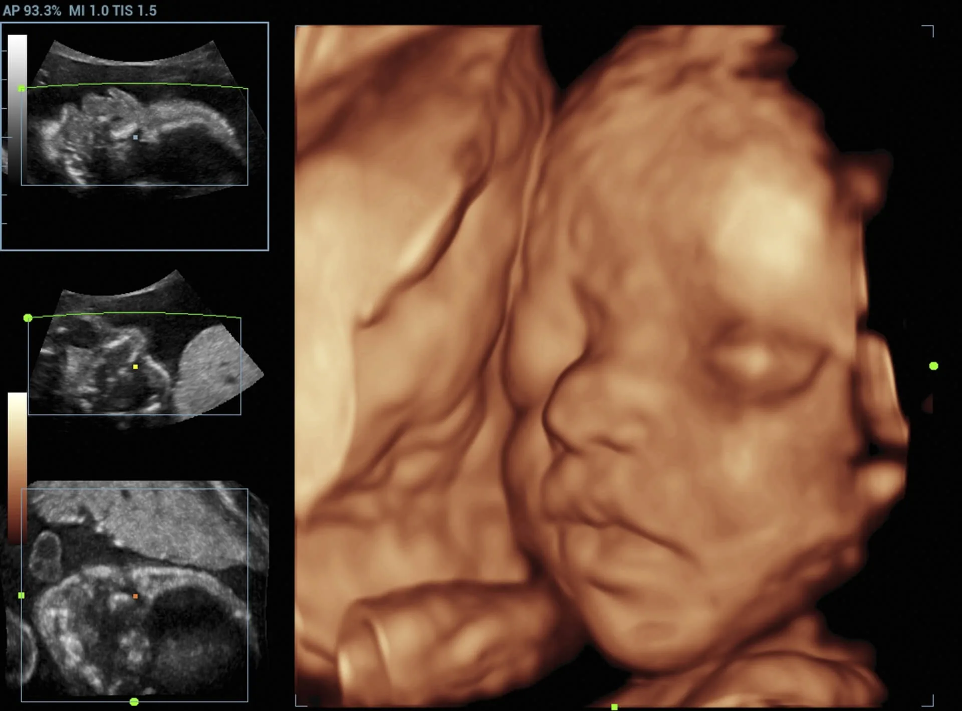
The anatomy scan — also called the mid-trimester survey or “20-week ultrasound” — is the most comprehensive ultrasound of pregnancy. It is a systematic evaluation of fetal anatomy including the brain, face, spine, heart (four-chamber view and outflow tracts), kidneys, bladder, stomach, limbs, and umbilical cord. Placental location, amniotic fluid volume, and cervical length are also assessed. This is typically when fetal sex can be determined if desired. The anatomy scan takes 30 to 45 minutes.
Serial growth ultrasounds are performed when there is concern about fetal growth — either too small (intrauterine growth restriction) or too large (macrosomia). These are typically ordered every 2 to 4 weeks and include measurements of the head, abdomen, and femur to calculate estimated fetal weight. Growth monitoring is standard in high-risk pregnancies including preeclampsia, gestational diabetes, multiple gestations, and chronic hypertension.
Third trimester ultrasounds may be performed to assess fetal position (particularly if breech presentation is suspected near term), amniotic fluid volume, placental location (confirming a low-lying placenta has migrated), and fetal well-being. Not every patient requires a third trimester ultrasound, but they are common in the later weeks of pregnancy.
For a complete breakdown of the prenatal screening schedule, see our Prenatal Care Guide.
Ultrasound is not exclusively a pregnancy tool. Gynecologic ultrasound is a fundamental part of evaluating a wide range of conditions outside of pregnancy. At Broad Medical Group, pelvic ultrasound is performed in-office for:
Gynecologic ultrasound is typically performed transvaginally, which provides higher-resolution images of the uterus and ovaries compared to the transabdominal approach. The exam is brief (usually 10 to 15 minutes) and results are reviewed with you immediately.
In addition to diagnostic imaging, Broad Medical Group offers elective 3D and 4D ultrasound sessions for parents who want to see their baby in greater detail. These sessions provide stunning surface-rendered images and real-time video — see your baby yawning, stretching, sucking their thumb, and making facial expressions.
The optimal window for elective 3D/4D imaging is between 26 and 32 weeks of pregnancy. At this stage:
Earlier scans (20–25 weeks) can still produce good images but facial features appear less filled-out. Later scans (33+ weeks) may be limited by the baby’s larger size and tighter quarters.
Several factors influence the clarity of 3D and 4D images:
If conditions are not ideal on the day of your appointment, Dr. Broad will still obtain the best images possible and may recommend rescheduling if visualization is significantly limited.
Schedule your 3D/4D session between 26 and 32 weeks for the best images. The sweet spot is around 28 weeks. Drink plenty of water in the days leading up to your appointment — good hydration supports amniotic fluid levels. 3D/4D sessions can be added to a scheduled prenatal visit or booked as a standalone appointment.
3D/4D ultrasound is not a substitute for diagnostic imaging. Elective 3D/4D sessions are a wonderful bonding experience, but they do not replace the standard 2D anatomy scan. The anatomy scan at 18–22 weeks provides the systematic structural evaluation needed to assess fetal development. 3D/4D is a complement, not a replacement.
For some transabdominal ultrasounds — particularly early pregnancy scans — you may be asked to arrive with a full bladder. A full bladder pushes the uterus into a better position for imaging and creates an acoustic window that improves image quality. You will be instructed on preparation when scheduling. For transvaginal scans and most second/third trimester scans, no special preparation is needed.
You will lie on an exam table. For transabdominal ultrasound, warm gel is applied to your abdomen and a handheld transducer is moved across the surface. For transvaginal ultrasound, a slim, lubricated probe is gently inserted into the vaginal canal. Neither approach is painful, though transvaginal ultrasound may cause mild pressure.
Scan duration varies by type:
At Broad Medical Group, Dr. Broad reviews results with you in real time during your appointment. You will see the images on the screen as they are captured, and Dr. Broad will explain what is being evaluated and what the findings show. There is no waiting for results to come back from an outside facility — this is a significant advantage of in-office imaging.
Diagnostic ultrasound uses sound waves — not radiation. It has been used in obstetrics for over 50 years with no confirmed adverse effects on mother or baby. Both ACOG and the American Institute of Ultrasound in Medicine (AIUM) endorse the safety of diagnostic ultrasound when performed by qualified providers for clinical indications. The ALARA principle (As Low As Reasonably Achievable) guides all imaging — using the minimum exposure needed for diagnostic purposes.
Whether you need a diagnostic scan or want to see your baby in 3D, Broad Medical Group provides in-office ultrasound with same-visit results. Dr. Broad is accepting new patients in Newport Beach and Orange County.
Book an Ultrasound →